Education9 min readAuthorMass Loaded Vinyl DirectPublishedUpdated

    How Does the Human Ear Convert Sound Into What We Hear?

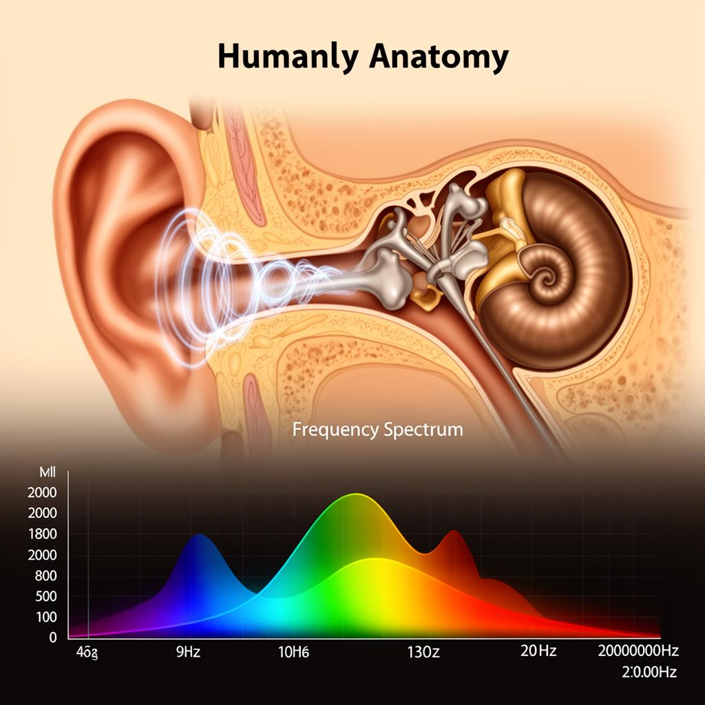
    Anatomical cross-section of the human ear showing the eardrum, ossicles, cochlea, and auditory nerve pathway illustrating how sound becomes hearing
    Anatomical cross-section of the human ear showing the eardrum, ossicles, cochlea, and auditory nerve pathway illustrating how sound becomes hearing

    1The Journey of a Sound Wave

    Before we dive into anatomy, here's the big picture. Sound travels through the ear in a four-stage relay system, with each stage converting energy from one form to another:
    Stage 1 — Acoustic energy (air vibrations) enters the outer ear and travels down the ear canal to the eardrum.
    Stage 2 — Mechanical energy (physical vibration) is created when the eardrum vibrates and transfers motion through three tiny bones in the middle ear.
    Stage 3 — Hydraulic energy (fluid waves) is generated when the last bone pushes against a fluid-filled structure called the cochlea in the inner ear.
    Stage 4 — Electrical energy (nerve impulses) is produced when specialized hair cells inside the cochlea convert fluid motion into electrical signals sent to the brain via the auditory nerve.
    This acoustic → mechanical → hydraulic → electrical conversion chain is one of the most elegant signal-processing systems in nature. Each stage amplifies, filters, and refines the signal so your brain receives precise information about frequency, amplitude, and timing.

    2The Outer Ear: Catching and Funneling Sound

    The outer ear consists of two parts: the pinna (the visible cartilage structure on the side of your head) and the ear canal (auditory meatus), a tube roughly 2.5 cm (1 inch) long that leads to the eardrum.

    The Pinna: Nature's Acoustic Funnel

    The pinna's complex folds and ridges aren't decorative — they're acoustic engineering. The curved shape collects sound waves from a wide area and funnels them into the narrow ear canal, providing a modest gain of about 5–6 dB. More importantly, the pinna's asymmetric shape creates tiny time delays and frequency-dependent filtering that help your brain determine whether a sound is coming from above, below, in front, or behind you. This is called spectral shaping, and it's unique to each person's ear shape.

    The Ear Canal: A Resonant Tube

    The ear canal acts as a resonant tube that naturally amplifies frequencies around 2,000–5,000 Hz by approximately 10–15 dB. This is not a coincidence — this frequency range corresponds to the most important frequencies in human speech. Evolution has literally tuned our ear canals to boost the sounds most critical for communication and survival.
    The ear canal also protects the delicate eardrum from physical damage, temperature extremes, and debris. Earwax (cerumen) traps dust and insects while keeping the canal lubricated.

    3The Middle Ear: The World's Smallest Amplifier

    At the end of the ear canal sits the tympanic membrane — the eardrum. This paper-thin membrane (about 0.1 mm thick and 8–10 mm in diameter) vibrates in response to incoming sound pressure waves. Despite its delicacy, the eardrum can detect pressure variations as small as 20 micropascals — displacement smaller than the diameter of a hydrogen atom.

    The Ossicles: Three Bones That Changed Everything

    Behind the eardrum lies a small air-filled cavity containing the three smallest bones in the human body — the ossicles:
    Malleus (Hammer): Attached directly to the eardrum, it picks up vibrations from the membrane and transfers them to the next bone.
    Incus (Anvil): Connected to the malleus, it acts as a bridge, transferring and refining the mechanical vibration.
    Stapes (Stirrup): The smallest bone in the human body (about 3 mm tall), it presses against the oval window of the cochlea, delivering concentrated mechanical force to the inner ear fluid.

    Impedance Matching: Solving the Air-to-Fluid Problem

    Here's the engineering challenge: sound travels efficiently through air, but the inner ear is filled with fluid. When sound hits a fluid boundary directly, about 99.9% of the energy is reflected — only 0.1% gets through. That's a 30 dB loss, which would make you nearly deaf.
    The middle ear solves this with an ingenious amplification system. The eardrum's surface area is about 17 times larger than the stapes footplate pressing on the oval window. This area ratio concentrates the force, and the lever action of the ossicle chain adds another 1.3× gain. Together, these mechanisms provide approximately 25–30 dB of amplification — almost perfectly compensating for the air-to-fluid energy loss. Engineers call this impedance matching, and the middle ear does it with three bones smaller than a grain of rice.

    The Acoustic Reflex: Built-In Protection

    Two tiny muscles attached to the ossicles — the tensor tympani and stapedius — contract reflexively when exposed to loud sounds (above ~85 dB). This stiffens the ossicle chain, reducing the energy transmitted to the inner ear by about 10–15 dB. However, this reflex has a latency of about 25–150 milliseconds, so it cannot protect against sudden impulse sounds like gunshots or explosions.

    4The Inner Ear: Where Sound Becomes Electricity

    The inner ear contains the cochlea — a snail-shaped, fluid-filled structure about the size of a pea that coils 2.5 turns. Despite its tiny size, the cochlea is the most complex mechanical structure in the human body and the true marvel of the hearing system.

    The Basilar Membrane: Nature's Frequency Analyzer

    Running the length of the cochlea is the basilar membrane, a thin ribbon of tissue that varies in width and stiffness along its length. At the base (near the oval window), it's narrow and stiff, responding to high frequencies (up to 20,000 Hz). At the apex (the tip of the spiral), it's wide and flexible, responding to low frequencies (down to 20 Hz).
    When the stapes pushes on the oval window, it creates a pressure wave in the cochlear fluid. This wave travels along the basilar membrane, and the membrane vibrates most strongly at the location corresponding to the wave's frequency. A 4,000 Hz tone causes peak vibration near the base; a 200 Hz tone causes peak vibration near the apex. This spatial mapping of frequency to position is called tonotopic organization, and it's how the ear performs real-time frequency analysis — essentially a biological Fourier transform.

    Hair Cells: The Biological Microphones

    Sitting on top of the basilar membrane are approximately 15,000–16,000 hair cells arranged in rows. Each hair cell has a bundle of tiny hair-like projections called stereocilia on its top surface. When the basilar membrane vibrates at a particular location, the stereocilia on the hair cells at that location bend.
    This bending opens mechanically-gated ion channels — tiny protein doors in the cell membrane. Potassium and calcium ions rush in, creating an electrical voltage change (depolarization) that triggers the release of neurotransmitters at the base of the hair cell. These neurotransmitters stimulate the fibers of the auditory nerve.
    There are two types of hair cells: Inner hair cells (IHCs) — about 3,500 cells arranged in a single row — are the primary sound sensors. They are responsible for converting mechanical vibration into the nerve signals your brain interprets as sound. Outer hair cells (OHCs) — about 12,000 cells in three rows — are biological amplifiers. They actively contract and expand in response to sound, amplifying quiet sounds by up to 40–60 dB and sharpening the frequency selectivity of the basilar membrane. This active amplification is why you can hear a whisper across a quiet room.
    Critical fact: Humans are born with a fixed number of hair cells, and they do not regenerate. Every hair cell lost to noise damage, aging, or toxins is gone permanently.

    5The Auditory Nerve: Delivering the Signal

    The auditory nerve (cochlear nerve) consists of approximately 30,000 nerve fibers bundled together, carrying electrical impulses from the hair cells to the brainstem. Each nerve fiber connects to a specific region of the cochlea, maintaining the tonotopic organization — high-frequency fibers from the base, low-frequency fibers from the apex.
    The auditory nerve encodes three critical pieces of information: Frequency is encoded by which fibers are firing (place coding) and by the timing pattern of nerve impulses (temporal coding, effective up to about 4,000 Hz). Loudness is encoded by the firing rate of individual fibers and by the number of fibers activated — louder sounds recruit more fibers. Timing is encoded with remarkable precision — the auditory nerve can resolve time differences as small as 10 microseconds, which is how your brain determines the direction a sound is coming from.
    The signal travels from the auditory nerve to the cochlear nucleus in the brainstem, then through several relay stations including the superior olivary complex (where signals from both ears are first compared for directional processing), the inferior colliculus, and the medial geniculate body before reaching the auditory cortex in the temporal lobe of the brain.

    6How the Brain Interprets Sound

    The auditory cortex doesn't just receive raw frequency and volume data — it performs extraordinarily complex processing to create your conscious experience of sound:
    Pattern recognition: Your brain identifies familiar sounds (a door closing, a dog barking, a specific person's voice) by matching incoming patterns against stored acoustic memories. This happens in milliseconds.
    Speech processing: Specialized areas like Wernicke's area decode the rapid frequency and timing patterns of speech into words and meaning. The brain can parse speech at rates up to 400 words per minute.
    Spatial processing: By comparing the tiny differences in arrival time and volume between your two ears (interaural time difference and interaural level difference), your brain constructs a 3D sound map of your environment. You can localize a sound source to within about 1–2 degrees horizontally.
    Emotional processing: The auditory cortex connects directly to the amygdala (emotional processing center), which is why certain sounds — a baby crying, a sudden loud bang, a loved one's voice — trigger immediate emotional responses before you consciously process what you heard.
    Selective attention: Perhaps most impressively, your brain can focus on one sound source while suppressing others — the cocktail party effect. In a noisy room with dozens of simultaneous conversations, you can selectively listen to one person while filtering out everything else.

    7Why Hearing Damage Is Irreversible

    Understanding the ear's anatomy explains why noise-induced hearing loss is permanent and why soundproofing matters for health:
    Hair cells don't regenerate. Unlike skin cells or blood cells, the hair cells in the cochlea are not replaced when damaged. Birds and some reptiles can regenerate hair cells, but mammals — including humans — cannot. Once a hair cell dies, the frequencies it was responsible for detecting are lost forever.
    Loud sounds physically destroy hair cells. Excessive sound pressure causes the stereocilia to bend beyond their elastic limit, breaking them off or fusing them together. Sustained exposure above 85 dB (the level of heavy traffic or a loud restaurant) begins damaging outer hair cells within hours. Exposure above 120 dB (a rock concert at the front row) can cause immediate, permanent damage.
    The damage is cumulative. Noise exposure acts like a lifetime damage meter. Every episode of excessive noise kills some hair cells, and the loss accumulates over years. This is why construction workers, musicians, factory workers, and anyone in consistently loud environments experience progressive hearing loss — even if no single exposure seemed dangerous at the time.
    High frequencies go first. Because the high-frequency hair cells are located at the base of the cochlea — right where sound energy enters — they receive the most intense vibration and are damaged first. This is why age-related and noise-induced hearing loss typically begins with difficulty hearing high-pitched sounds (consonants like 's', 'f', 'th') while low-frequency hearing remains relatively intact.
    The World Health Organization estimates that 1.1 billion young people are at risk of hearing loss from unsafe listening practices, making acoustic protection and soundproofing not just a comfort issue but a public health priority.

    8Connections to Soundproofing and Acoustics

    The biology of hearing directly informs practical soundproofing decisions:

    Why STC Ratings Exist

    Sound Transmission Class (STC) ratings weight different frequencies based on human hearing sensitivity — the same sensitivity curve shaped by the ear canal's resonance and the cochlea's tonotopic mapping. An STC 50 wall blocks more mid-frequency energy because that's where human hearing (and speech) is most sensitive.

    Why Low-Frequency Noise Is So Problematic

    Bass frequencies require more mass to block because low-frequency sound waves carry more energy per cycle. But the ear's biology adds another dimension: low-frequency sounds are detected by hair cells at the apex of the cochlea, in a region that's harder to protect with the acoustic reflex. Prolonged bass exposure can cause damage that's not immediately apparent because it doesn't trigger the same "this is too loud" sensation as mid-frequency noise.

    Why Mass Loaded Vinyl Works With Your Hearing System

    Mass loaded vinyl (MLV) is effective across the frequency range most critical to human hearing — roughly 250–6,000 Hz. This range encompasses speech intelligibility, most musical content, and the frequencies where the ear is most sensitive. By adding mass to wall and floor assemblies, MLV reduces the sound energy reaching your ears, directly protecting the irreplaceable hair cells in your cochlea.

    10Conclusion

    The human ear is one of nature's most extraordinary instruments — a system that converts invisible air vibrations into the rich auditory world you experience every moment of every day. From the pinna's acoustic funneling to the cochlea's biological frequency analysis to the brain's pattern recognition, each stage is optimized for sensitivity, speed, and precision. Understanding this system makes the case for hearing protection and proper soundproofing unmistakably clear: the components that make hearing possible are irreplaceable, and every decibel of unnecessary noise exposure brings permanent damage one step closer. Whether you're designing a quiet office, soundproofing a bedroom, or simply choosing when to wear ear protection, you're making decisions that directly affect the 15,000 hair cells standing between you and silence.

    FAQs: How Does the Human Ear Work

    Need Expert Soundproofing Advice?

    Our team of soundproofing specialists is here to help you choose the right products and installation methods for your museum or gallery project.